Pubblicazioni

iForensic, multicentric validation of digital whole slide images (WSI) in forensic histopathology setting according to the College of American Pathologists guidelines  (2025)

Autori:
Pigaiani, Nicola; Oliva, Antonio; Cirielli, Vito; Grassi, Simone; Arena, Vincenzo; Solari, Luca-Maria; Tatriele, Naomi; Raniero, Dario; Brunelli, Matteo; Gobbo, Stefano; Scarpa, Aldo; Pantanowitz, Liron; Rodegher, Pamela; Bortolotti, Federica; Ausania, Francesco
Titolo:
iForensic, multicentric validation of digital whole slide images (WSI) in forensic histopathology setting according to the College of American Pathologists guidelines
Anno:
2025
Tipologia prodotto:
Articolo in Rivista
Tipologia ANVUR:
Articolo su rivista
Lingua:
Inglese
Referee:
No
Nome rivista:
International Journal of Legal Medicine
ISSN Rivista:
0937-9827
N° Volume:
139
Numero o Fascicolo:
3
Intervallo pagine:
1161-1168
Parole chiave:
Digital forensic histopathology; Digital glass; Digital whole slide images; Forensic pathology; Method validation
Breve descrizione dei contenuti:
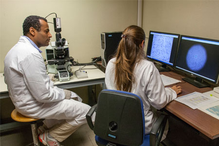
: Pathology has benefited from the rapid progress of image-digitizing technology during the last decade. However, the application of digital whole slide images (WSI) in forensic pathology still needs to be improved. WSI validation is crucial to ensure diagnostic performance, at least equivalent to glass slides and light microscopy. The College of American Pathologists Pathology and Laboratory Quality Center recently updated internal digital pathology system validation recommendations. Following these guidelines, this pilot study aimed to validate the performance of a digital approach for forensic histopathological diagnosis. Six independent skilled forensic pathologists from different forensic medicine institutes evaluated 100 glass slides of forensic interest (80 stained with standard hematoxylin and eosin, 20 with special staining), including different organs and tissues, with light microscopy (Olympus BX51, Tokyo, Japan). Glass slides were scanned using the Aperio GT 450 DX Digital Slides Scanner (Leica Biosystems, Nussloch, Germany). After two wash-out weeks, forensic pathologists evaluated WSIs in front of a widescreen using computer devices with dedicated software (O3 viewer, O3 Enterprise, Zucchetti, Trieste, Italy). Side-by-side comparisons between diagnoses performed on tissue glass slides versus WSIs were above the threshold stated in the validation guidelines (mean concordance of 97.8%). CSUQ Version 3 questionnaire showed high satisfaction for all pathologists (mean result: 6.6/7). Our institutional digital forensic pathology system has been validated for practical casework application. This approach opens new scenarios in practical forensic casework investigations, such as sharing live histological ex-glass slides online, as well as educational and research perspectives, with improving impacts on the whole daily workflow.
Note:
Epub 2025 Jan 21
Id prodotto:
144192
Handle IRIS:
11562/1153627
ultima modifica:
24 aprile 2025
Citazione bibliografica:
Pigaiani, Nicola; Oliva, Antonio; Cirielli, Vito; Grassi, Simone; Arena, Vincenzo; Solari, Luca-Maria; Tatriele, Naomi; Raniero, Dario; Brunelli, Matteo; Gobbo, Stefano; Scarpa, Aldo; Pantanowitz, Liron; Rodegher, Pamela; Bortolotti, Federica; Ausania, Francesco, iForensic, multicentric validation of digital whole slide images (WSI) in forensic histopathology setting according to the College of American Pathologists guidelines «International Journal of Legal Medicine» , vol. 139 , n. 32025pp. 1161-1168

Consulta la scheda completa presente nel repository istituzionale della Ricerca di Ateneo IRIS

<<indietro

Attività

Strutture

Condividi